The purpose of this project is to construct a Geiger counter to measure Pippa's radioactivity after a PET (Positron Emission Tomography) scan.
PET scans require the prior injection of a radioactive tracer which results in the patient being radioactive for a short period (a few hours).
The particular tracer used in this PET scan is fluorodeoxyglucose (FDG) F18 which contains a radioactive isotope of fluorine (fluorine 18) with a half-life of 1.83 hours.
This allows plenty of time for the scan while ensuring that the radioactivity disappears - naturally quite quickly.
Just before the PET scan Pippa was injected with FDG which is in effect radioactive glucose.
Any active cancerous tissue takes up the glucose more than normal tissue so the scan can build up a picture of what's going on inside the body from the emitted radiation. The radioactive isotope chosen (fluorine 18) only lasts for a few hours after creation so in the video we can see her radioactivity tailing off. Thankfully the results of the PET scan were good news!
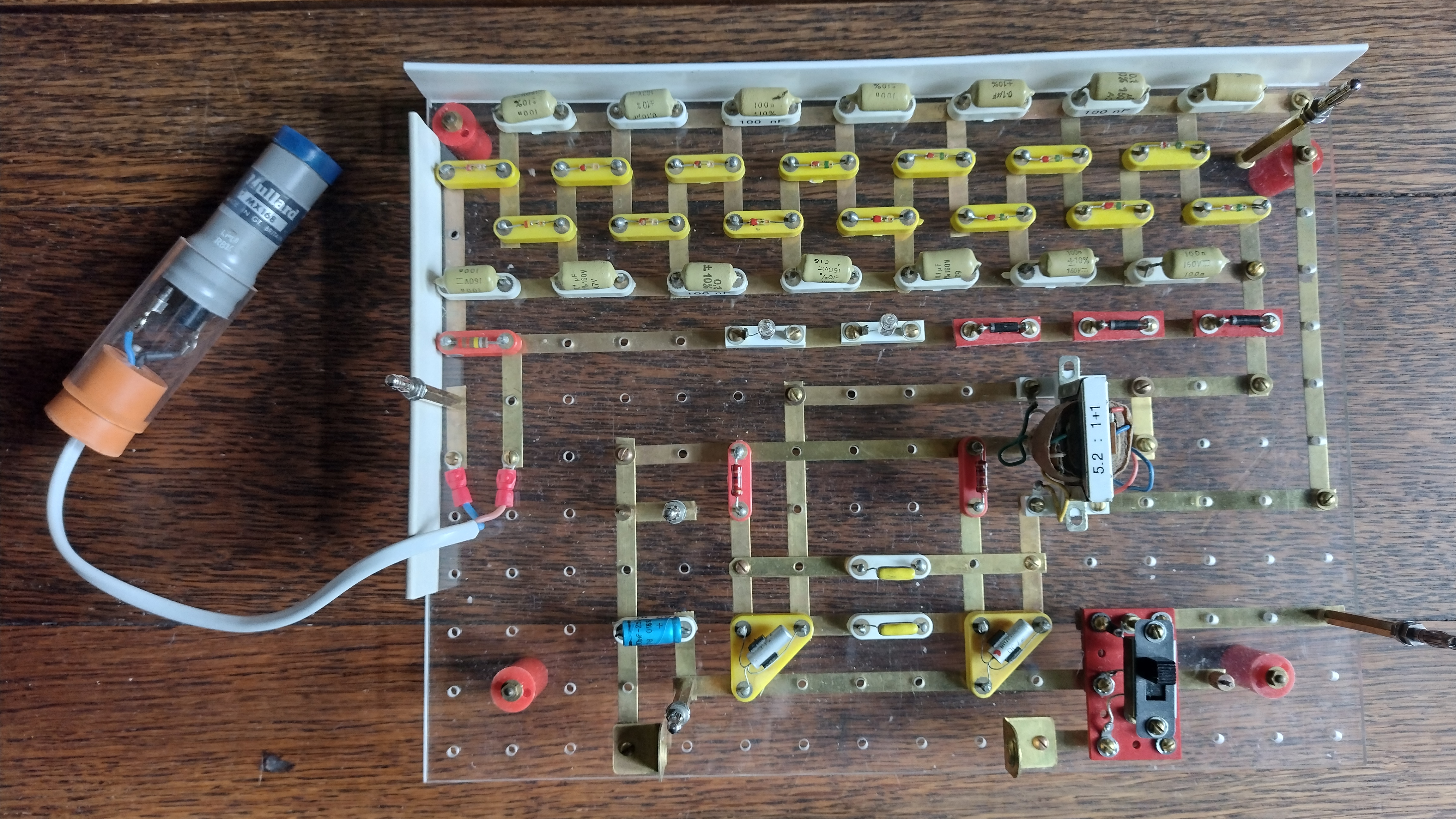
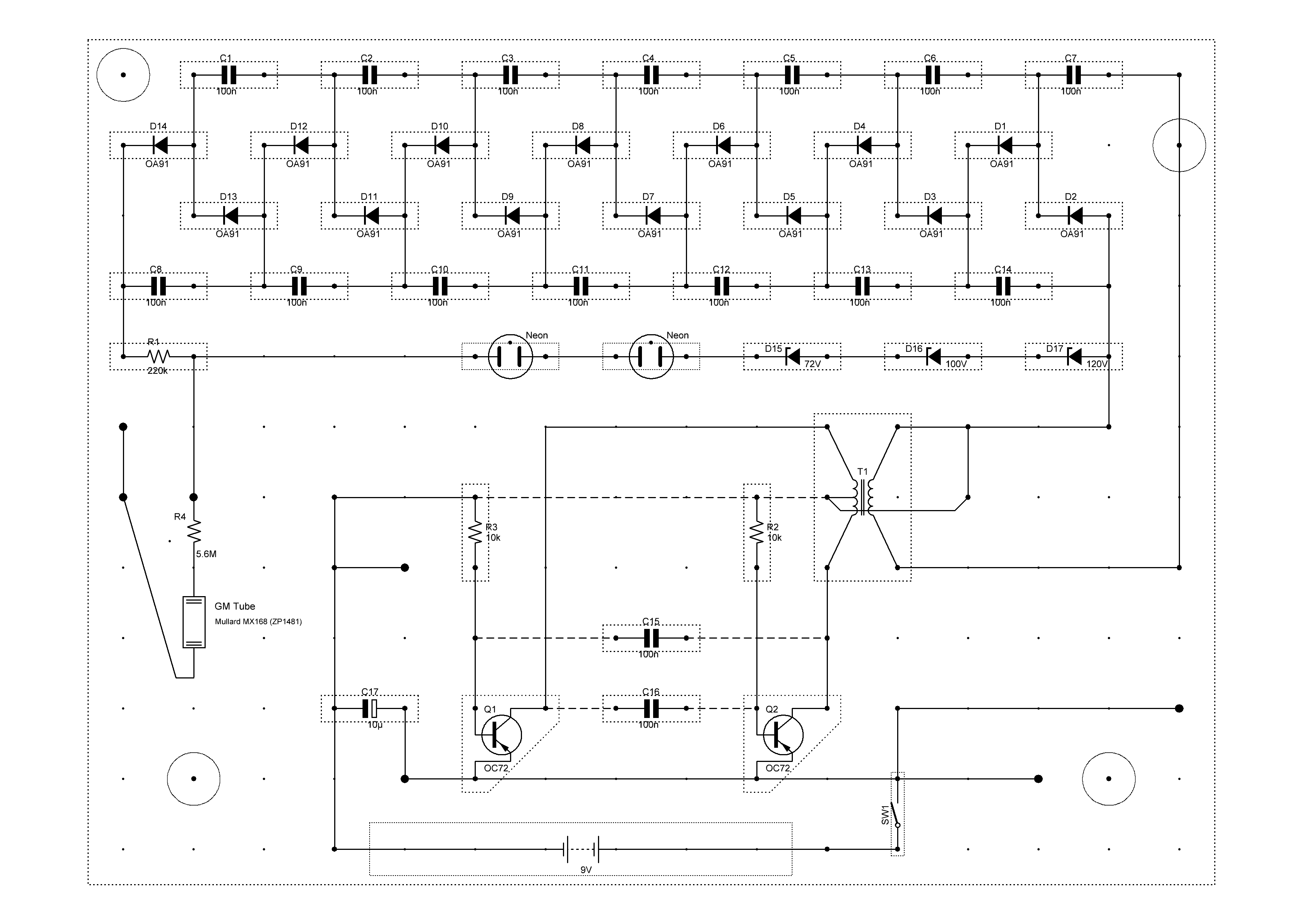
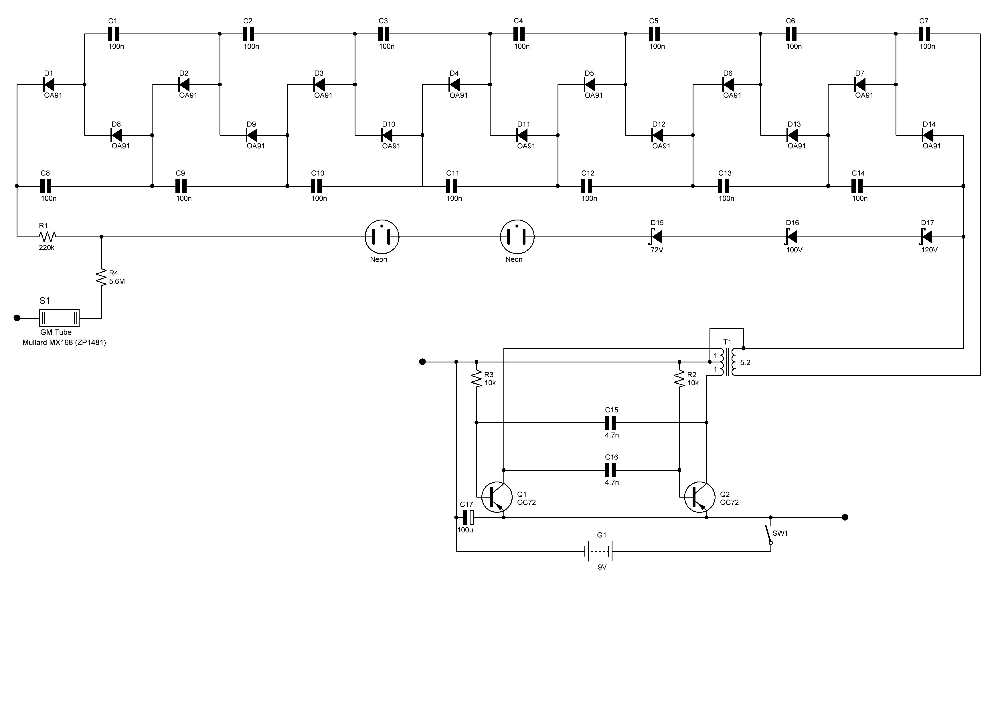
A Geiger-Müller tube consists of a chamber filled with a gas mixture at a low pressure (about 0.1 atmosphere).
The chamber contains two electrodes whose potentials differ by several hundred volts (400V in this case).
When ionising radiation strikes the tube, some molecules of the fill gas are ionised directly by the incident radiation.
The high potential difference inside the tube creates the right environment for the ionisation to trigger a 'Townsend avalanche',
causing a small current pulse of short duration across the tube.
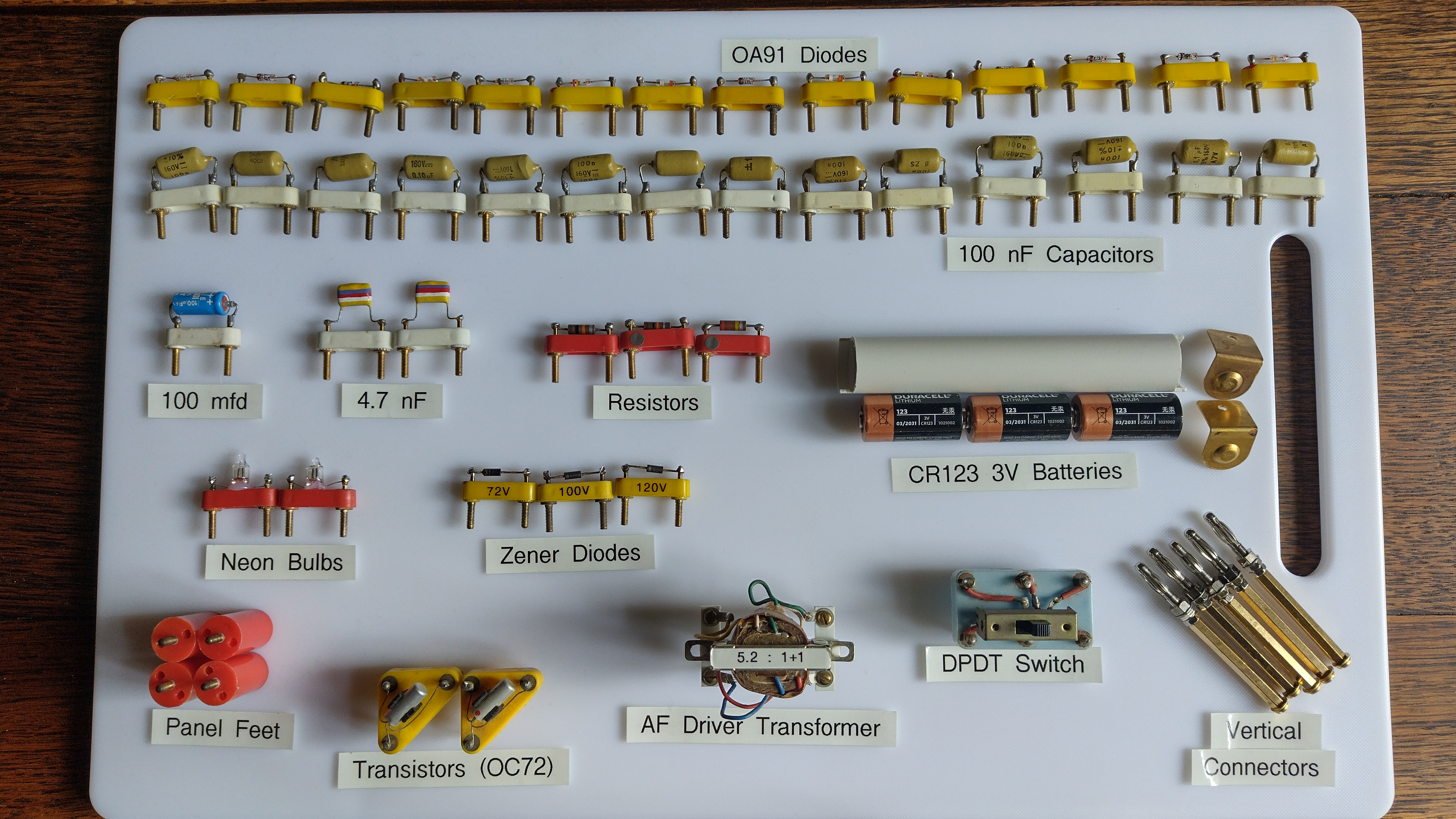
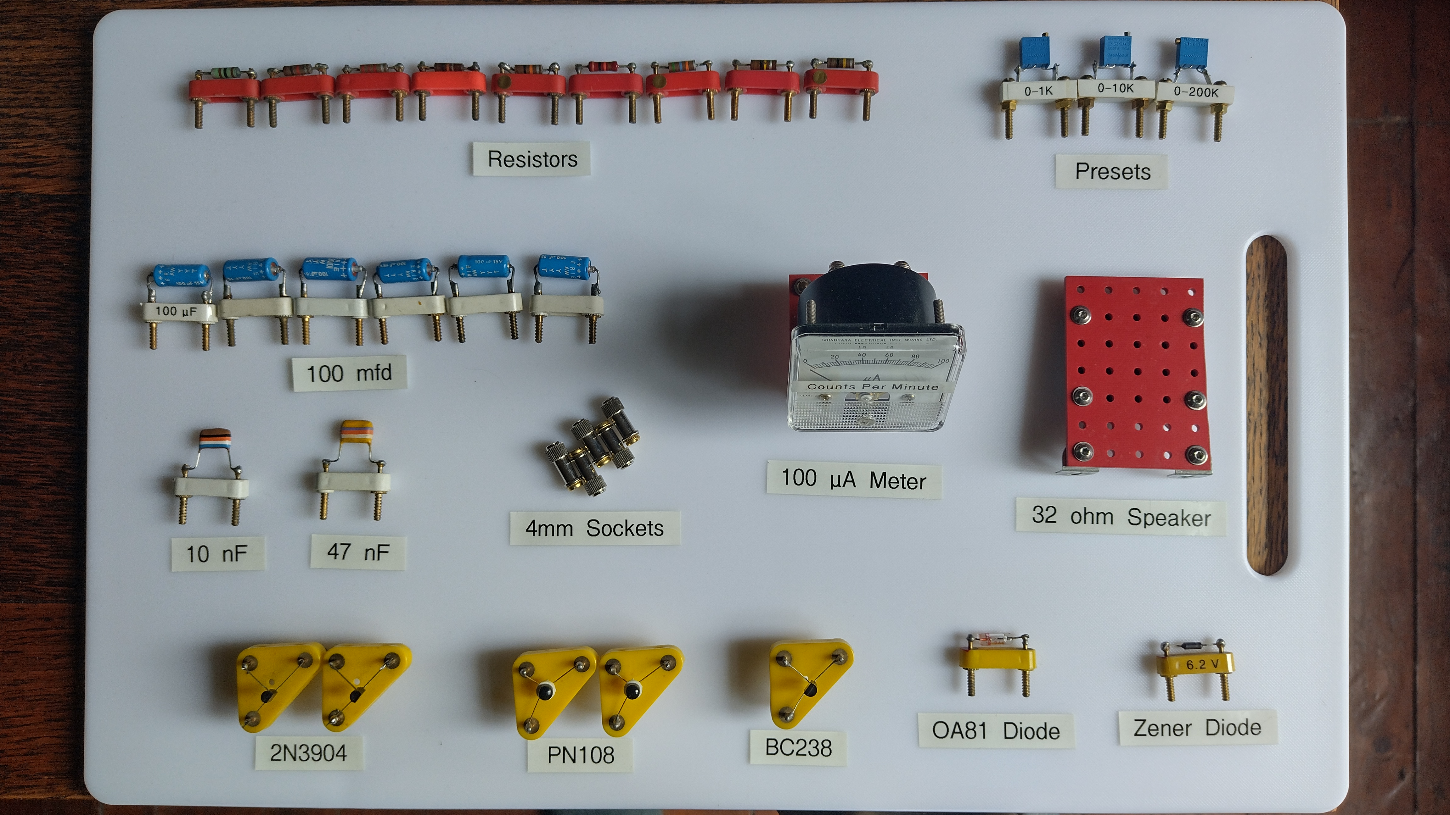
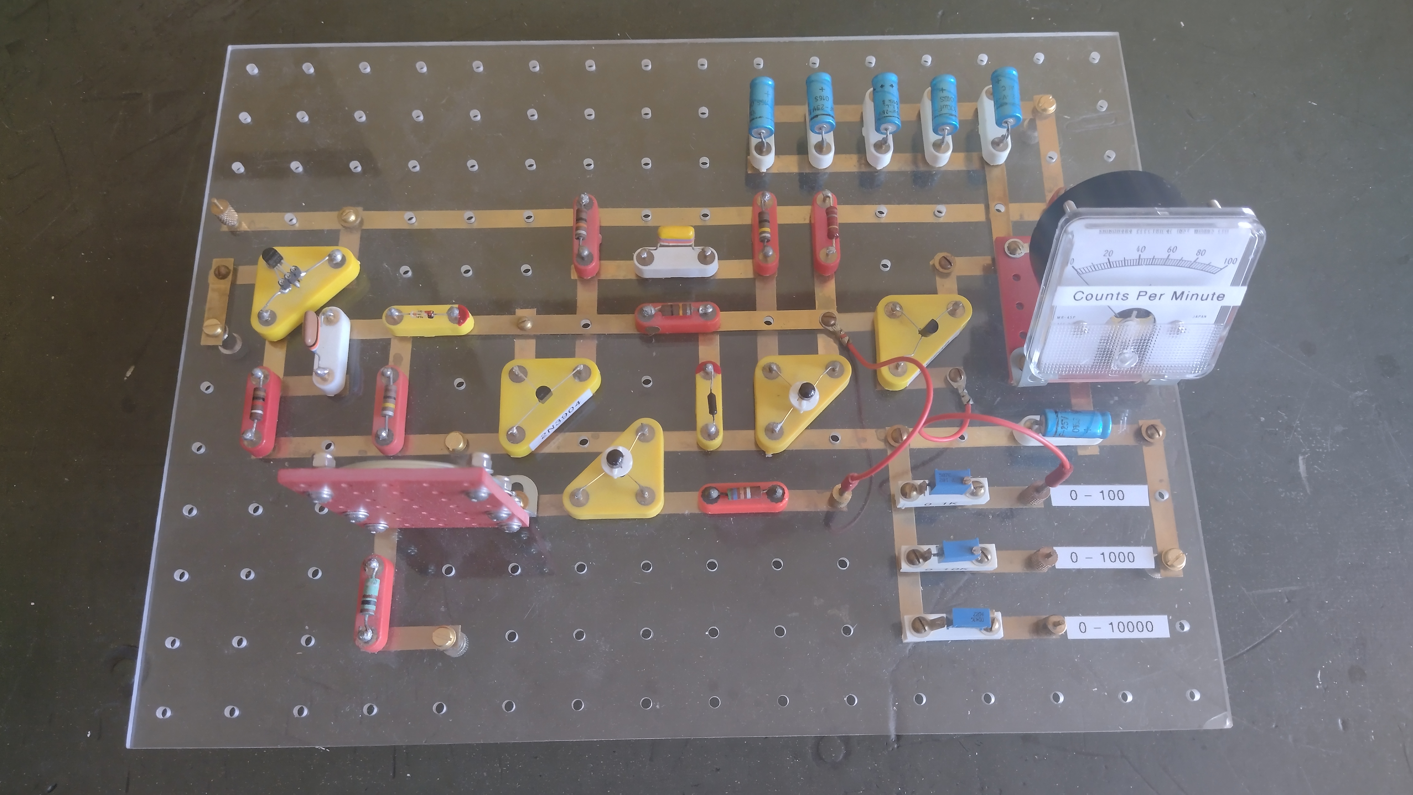
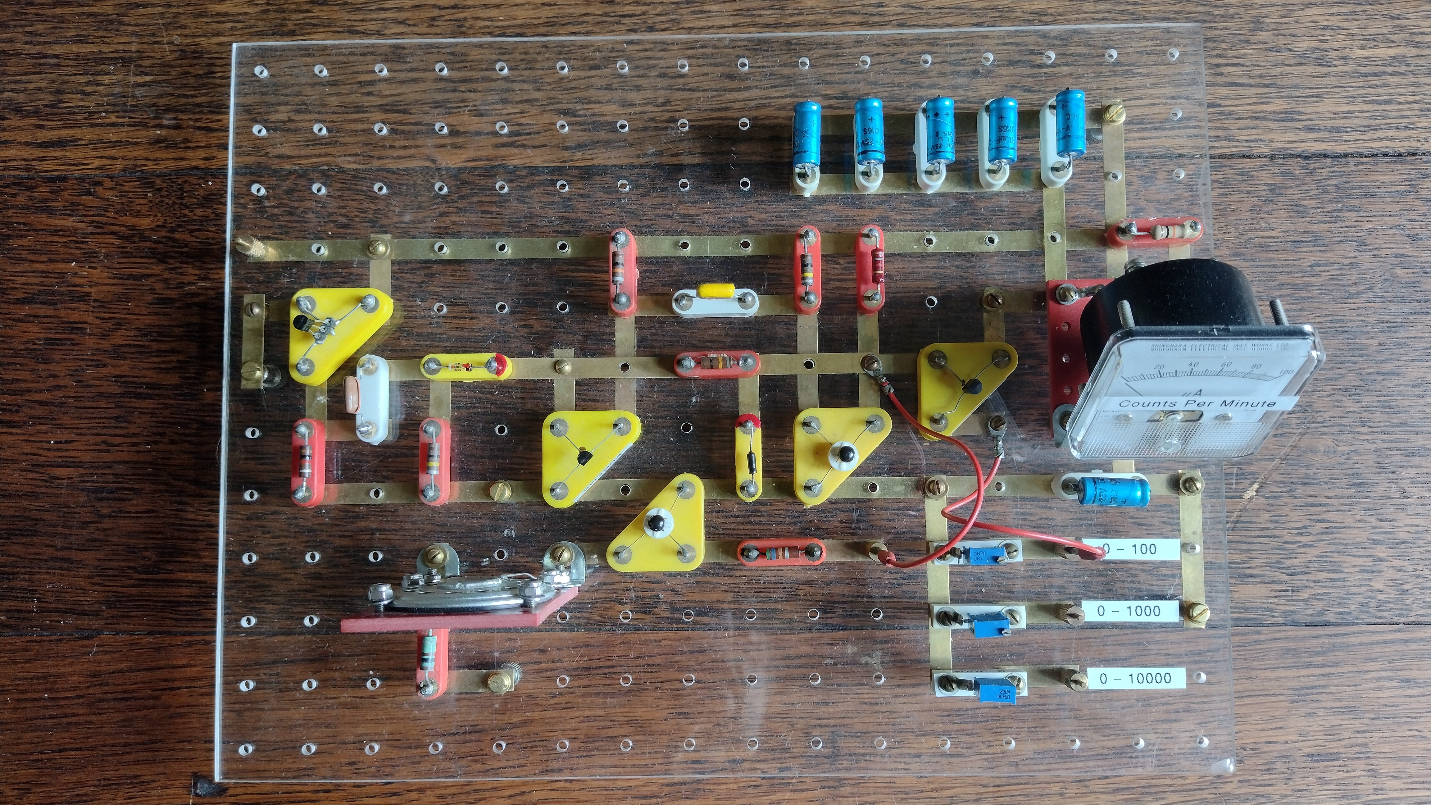
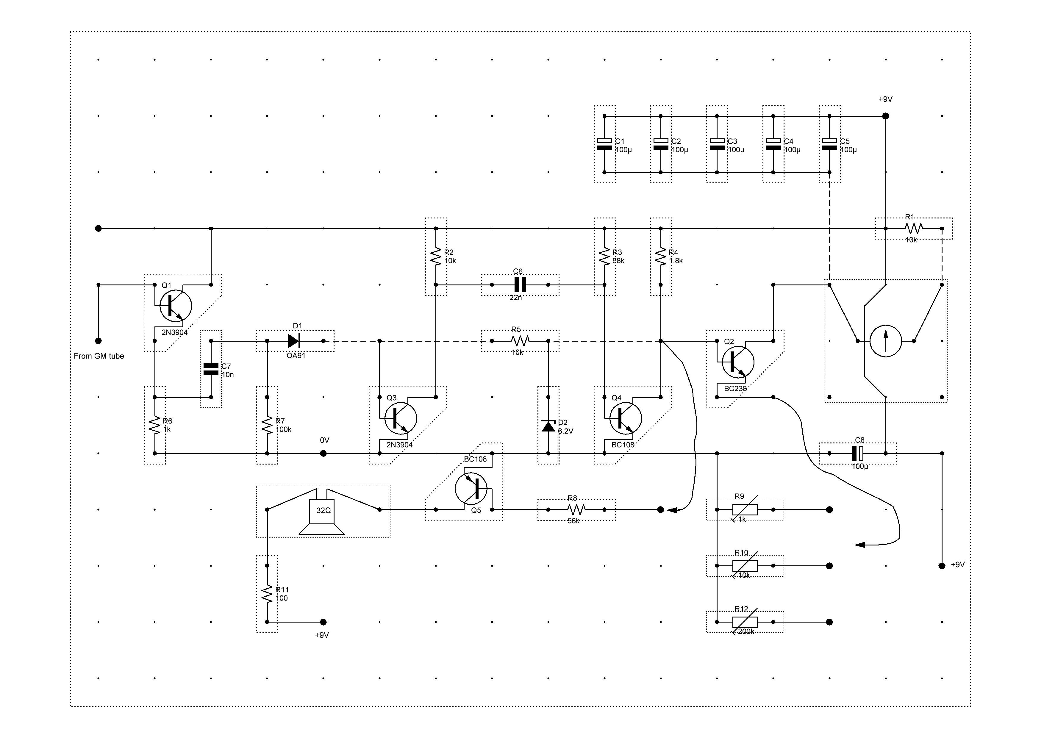
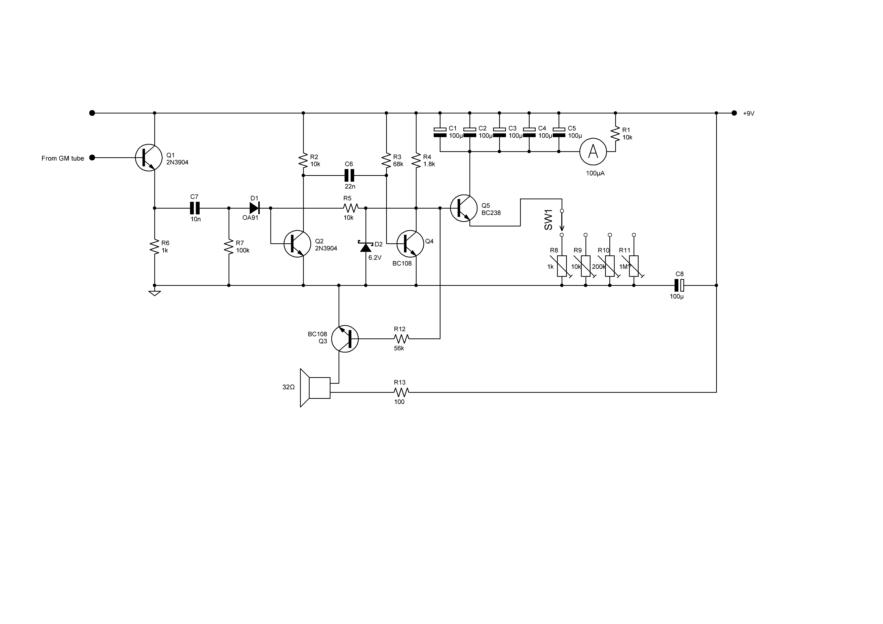
The Geiger-Müller tube in use in this project is the Mullard MX168 (ZP1481).
The gas mixture inside this tube is made up of neon, argon and a halogen. When detecting ionising radiation it produces 20 μA pulses of 100 μs duration.
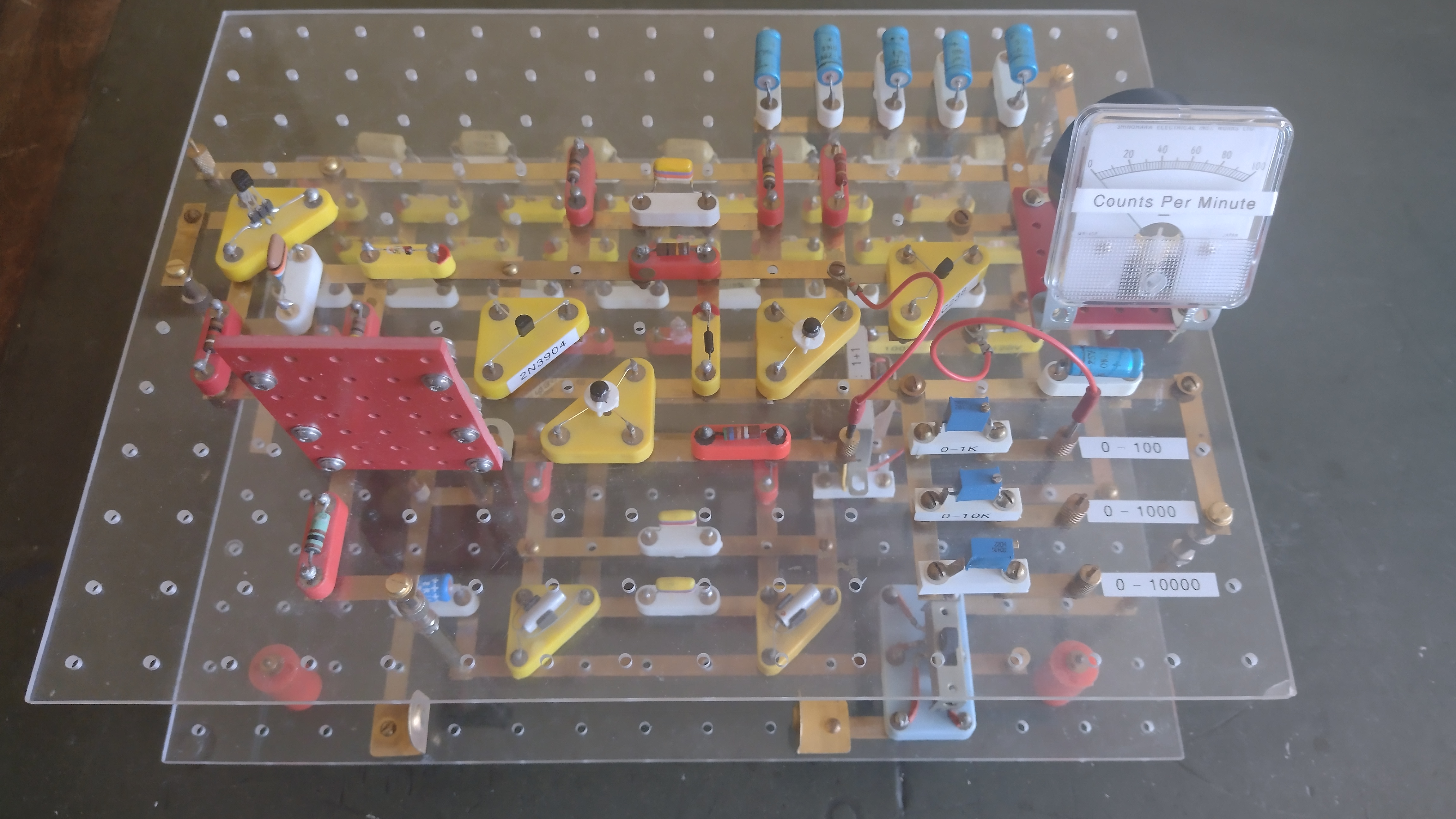
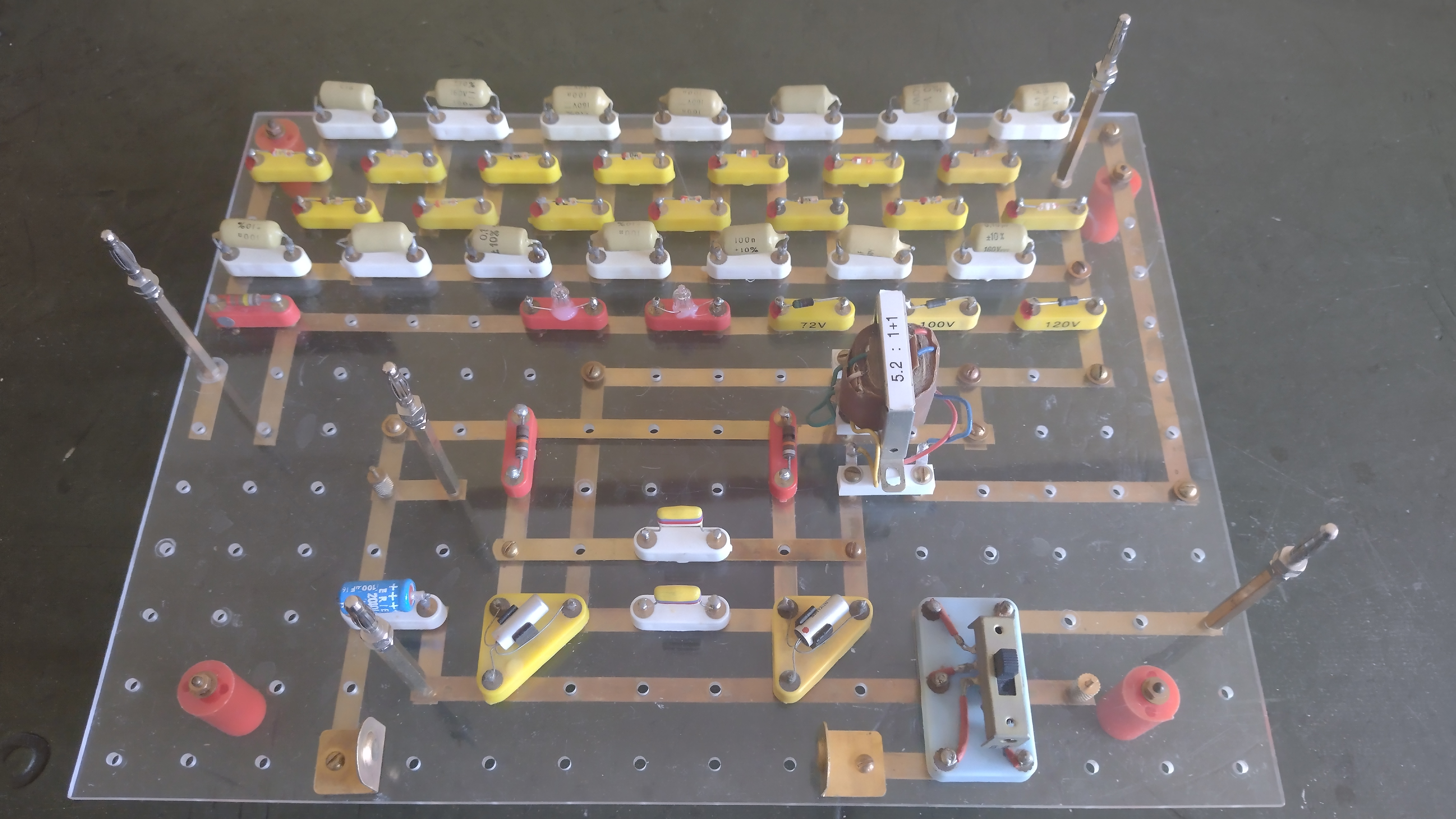
This Geiger counter has 2 decks:
* a bottom deck high voltage generator
* and a top deck pulse counter.